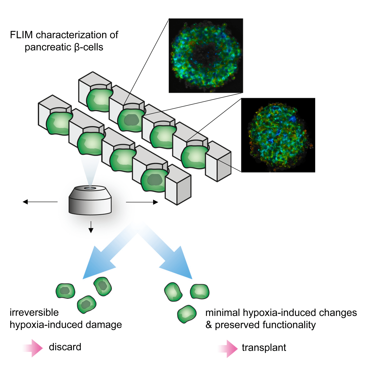
Fluorescence lifetime imaging microscopy (FLIM) is a powerful microscopy technique that can probe changes in metabolic state in vitro and in vivo in real-time, and can serve as diagnostic tools of pathological tissues in situ. The time-dimensional characteristic of FLIM combined with multiphoton (MP) microscopy enables the visualization of endogenous nicotinamide adenine dinucleotide (NADH) and flavin adenine dinucleotide (FAD), and the discrimination between free and protein-bound forms based on their respective lifetimes. In our work, we demonstrated how (pre-) hypoxic β-cell-composed pseudo-islets can be discriminated from healthy functional pseudo-islets based on their FLIM-based metabolic profiles. The use of FLIM during the pre-transplantation pancreatic islet selection process has the potential to improve the outcome of β-cell islet transplantation.
Fluorescence-lifetime imaging microscopy
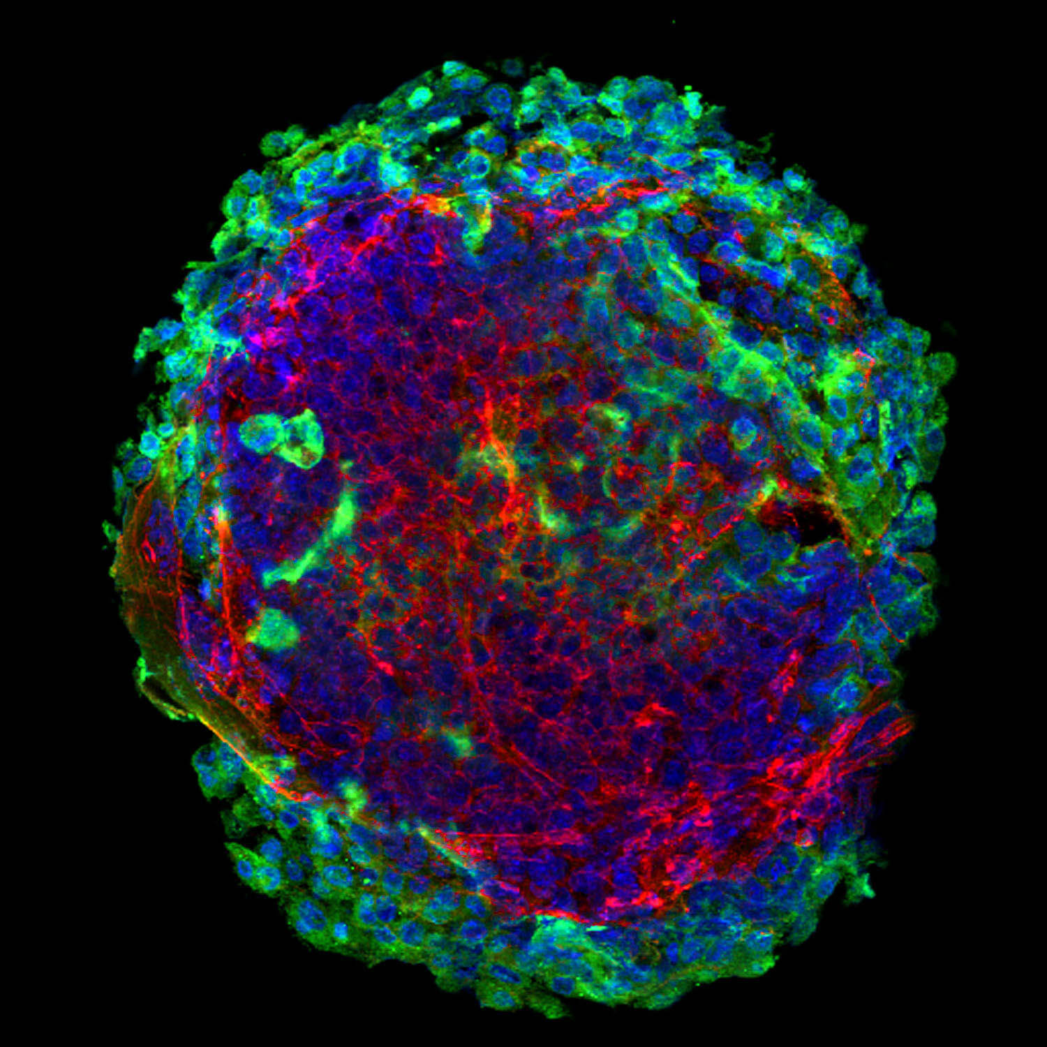
Confocal laser scanning microscopy
Confocal laser scanning has become a powerful method for the nondestructive evaluation of deep-tissue cells and extracellular matrix (ECM) structures. By interacting with highly non-centrosymmetric molecular assemblies, the non-linear process called second harmonic generation has also proven to be an important diagnostic tool for the visualization of ECM compartments in situ with submicron resolution without the need for tissue processing.